


Sodium, potassium, calcium, bicarbonate, and fluid balance in the blood are the basis for maintaining physiological functions in the body. There has been a lack of research on magnesium ion disorder. As early as the 1980s, magnesium was known as the “forgotten electrolyte”. With the discovery of magnesium specific channels and transporters, as well as the understanding of physiological and hormonal regulation of magnesium homeostasis, people’s understanding of the role of magnesium in clinical medicine is constantly deepening.
Magnesium is crucial for cellular function and health. Magnesium typically exists in the form of Mg2+, and is present in all cells of all organisms, from plants to higher mammals. Magnesium is an essential element for health and life, as it is an important cofactor of cellular energy source ATP. Magnesium mainly participates in the main physiological processes of cells by binding to nucleotides and regulating enzyme activity. All ATPase reactions require Mg2+- ATP, including reactions related to RNA and DNA functions. Magnesium is a cofactor of hundreds of enzymatic reactions in cells. In addition, magnesium also regulates glucose, lipid, and protein metabolism. Magnesium is involved in regulating neuromuscular function, regulating heart rhythm, vascular tone, hormone secretion, and the release of N-methyl-D-aspartate (NMDA) in the central nervous system. Magnesium is the second messenger involved in intracellular signaling and a regulator of circadian rhythm genes that control the circadian rhythm of biological systems.
There is approximately 25 g of magnesium in the human body, mainly stored in bones and soft tissues. Magnesium is an important intracellular ion and the second largest intracellular cation after potassium. In cells, 90% to 95% of magnesium binds to ligands such as ATP, ADP, citrate, proteins, and nucleic acids, while only 1% to 5% of intracellular magnesium exists in free form. The intracellular free magnesium concentration is 1.2-2.9 mg/dl (0.5-1.2 mmol/L), which is similar to the extracellular concentration. In plasma, 30% of circulating magnesium binds to proteins mainly through free fatty acids. Patients with long-term high levels of free fatty acids typically have lower blood magnesium concentrations, which are inversely proportional to the risk of cardiovascular and metabolic diseases. Changes in free fatty acids, as well as levels of EGF, insulin, and aldosterone, can affect blood magnesium levels.
There are three main regulatory organs of magnesium: the intestine (regulating dietary magnesium absorption), bones (storing magnesium in the form of hydroxyapatite), and kidneys (regulating urinary magnesium excretion). These systems are integrated and highly coordinated, together forming the gut bone kidney axis, responsible for the absorption, exchange, and excretion of magnesium. Imbalance of magnesium metabolism may lead to pathological and physiological outcomes
Foods rich in magnesium include grains, beans, nuts, and green vegetables (magnesium is the core component of chlorophyll). Approximately 30% to 40% of dietary magnesium intake is absorbed by the intestine. Most absorption occurs in the small intestine through intercellular transport, a passive process involving tight junctions between cells. The large intestine can finely regulate magnesium absorption through transcellular TRPM6 and TRPM7. The inactivation of the intestinal TRPM7 gene can lead to severe deficiencies in magnesium, zinc, and calcium, which is detrimental to early growth and survival after birth. Magnesium absorption is influenced by various factors, including magnesium intake, intestinal pH value, hormones (such as estrogen, insulin, EGF, FGF23, and parathyroid hormone [PTH]), and gut microbiota.
In the kidneys, renal tubules reabsorb magnesium through both extracellular and intracellular pathways. Unlike most ions such as sodium and calcium, only a small amount (20%) of magnesium is reabsorbed in the proximal tubules, while the majority (70%) of magnesium is reabsorbed in the Heinz loop. In the proximal tubules and the coarse branches of the Heinz loop, magnesium reabsorption is mainly driven by concentration gradients and membrane potential. Claudin 16 and Claudin 19 form magnesium channels in the thick branches of the Heinz loop, while Claudin 10b helps to form a positive intraluminal voltage across epithelial cells, driving magnesium ion reabsorption. In the distal tubules, magnesium finely regulates intracellular reabsorption (5%~10%) through TRPM6 and TRPM7 at the cell tip, thereby determining the final urinary magnesium excretion.
Magnesium is an important component of bones, and 60% of magnesium in the human body is stored in the bones. The exchangeable magnesium in bones provides dynamic reserves for maintaining plasma physiological concentrations. Magnesium promotes bone formation by affecting the activity of osteoblasts and osteoclasts. Increasing magnesium intake can increase bone mineral content, thereby reducing the risk of fractures and osteoporosis during aging. Magnesium has a dual role in bone repair. During the acute phase of inflammation, magnesium can promote the expression of TRPM7 in macrophages, magnesium dependent cytokine production, and promote the immune microenvironment of bone formation. During the late remodeling stage of bone healing, magnesium can affect osteogenesis and inhibit hydroxyapatite precipitation. TRPM7 and magnesium also participate in the process of vascular calcification by influencing the transition of vascular smooth muscle cells to osteogenic phenotype.
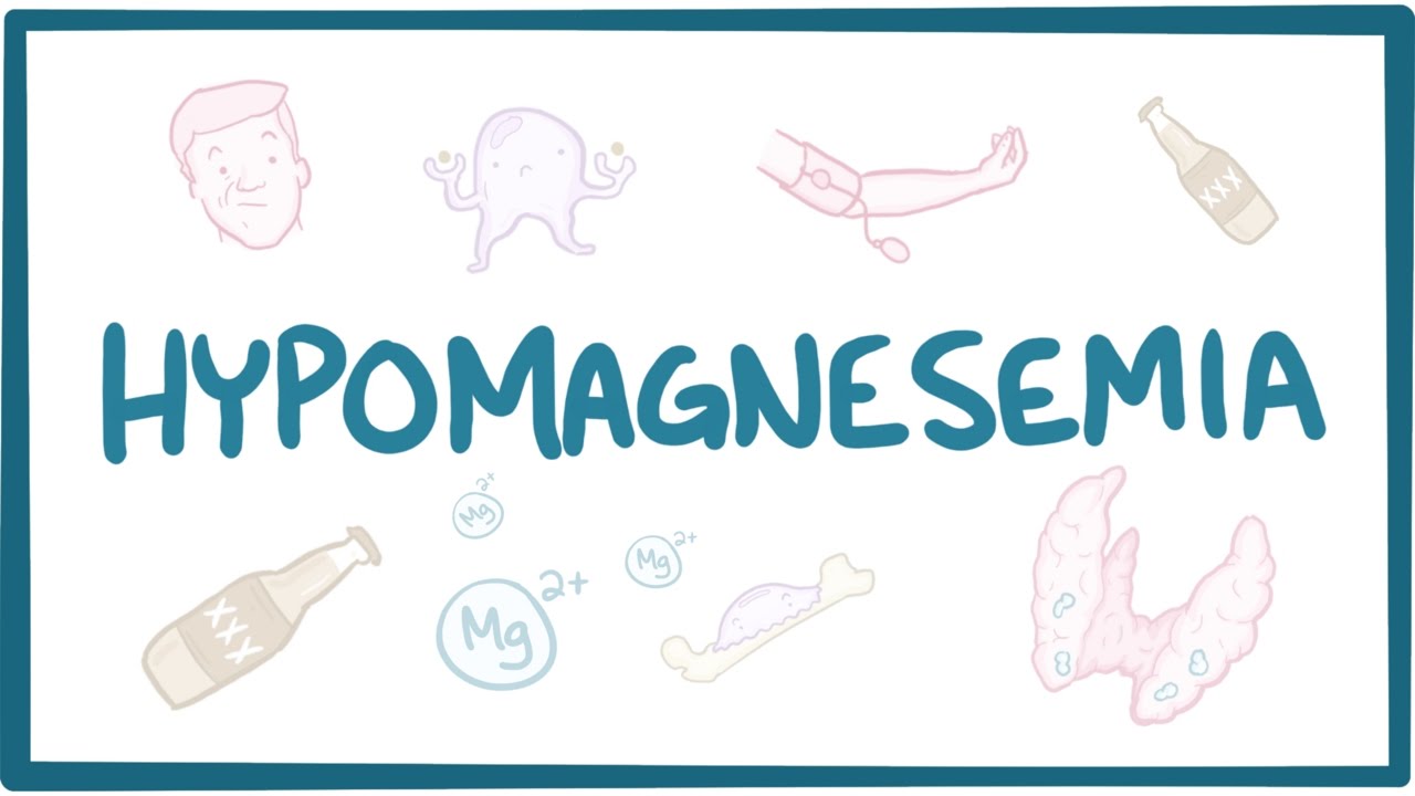
The normal serum magnesium concentration in adults is 1.7~2.4 mg/dl (0.7~1.0 mmol/L). Hypomagnesemia refers to a serum magnesium concentration below 1.7 mg/dl. Most patients with borderline hypomagnesemia have no obvious symptoms. Due to the possibility of long-term potential magnesium deficiency in patients with serum magnesium levels greater than 1.5 mg/dl (0.6 mmol/L), some suggest raising the lower threshold for hypomagnesemia. However, this level is still controversial and requires further clinical validation. 3%~10% of the general population has hypomagnesemia, while the incidence rate of type 2 diabetes patients (10%~30%) and hospitalized patients (10%~60%) is higher, especially in the intensive care unit (ICU) patients, whose incidence rate exceeds 65%. Multiple cohort studies have shown that hypomagnesemia is associated with an increased risk of all-cause mortality and cardiovascular disease-related mortality.
The clinical manifestations of hypomagnesemia include non-specific symptoms such as drowsiness, muscle spasms, or muscle weakness caused by insufficient dietary intake, increased gastrointestinal loss, reduced renal reabsorption, or redistribution of magnesium from the outside to the inside of cells (Figure 3B). Hypomagnesemia usually coexists with other electrolyte disorders, including hypocalcemia, hypokalemia, and metabolic alkalosis. Therefore, hypomagnesemia may be overlooked, especially in most clinical settings where blood magnesium levels are not routinely measured. Only in severe hypomagnesemia (serum magnesium<1.2 mg/dL [0.5 mmol/L]), symptoms such as abnormal neuromuscular excitability (wrist ankle spasms, epilepsy, and tremors), cardiovascular abnormalities (arrhythmias and vasoconstriction), and metabolic disorders (insulin resistance and cartilage calcification) become apparent. Hypomagnesemia is associated with increased hospitalization and mortality rates, especially when accompanied by hypokalemia, highlighting the clinical importance of magnesium.
The magnesium content in the blood accounts for less than 1%, so the blood magnesium content cannot reliably reflect the total magnesium content in the tissue. Research has shown that even if the serum magnesium concentration is normal, the intracellular magnesium content may be depleted. Therefore, considering only the magnesium content in the blood without considering dietary magnesium intake and urine loss may underestimate the clinical magnesium deficiency.
Patients with hypomagnesemia often experience hypokalemia. Stubborn hypokalemia is usually associated with magnesium deficiency, and it can only be effectively corrected after magnesium levels return to normal. Magnesium deficiency can promote potassium secretion from the collecting ducts, further exacerbating potassium loss. A decrease in intracellular magnesium levels inhibits Na+- K+- ATPase activity and increases the opening of extrarenal medullary potassium (ROMK) channels, leading to more potassium loss from the kidneys. The interaction between magnesium and potassium also involves activating the sodium chloride co transporter (NCC), thereby promoting sodium reabsorption. Magnesium deficiency reduces NCC abundance through an E3 ubiquitin protein ligase called NEDD4-2, which downregulates neuronal precursor cell development, and prevents NCC activation through hypokalemia. Continuous downregulation of NCC can enhance distal Na+transport in hypomagnesemia, leading to increased urinary potassium excretion and hypokalemia.
Hypocalcemia is also common in patients with hypomagnesemia. Magnesium deficiency can inhibit the release of parathyroid hormone (PTH) and reduce the sensitivity of the kidneys to PTH. A decrease in PTH levels can reduce renal calcium reabsorption, increase urinary calcium excretion, and ultimately lead to hypocalcemia. Due to hypocalcemia caused by hypomagnesemia, hypoparathyroidism is often difficult to correct unless blood magnesium levels return to normal.
Serum total magnesium measurement is the standard method for determining magnesium content in clinical practice. It can quickly assess short-term changes in magnesium content, but may underestimate the total magnesium content in the body. Endogenous factors (such as hypoalbuminemia) and exogenous factors (such as specimen hemolysis and anticoagulants, such as EDTA) may affect the measurement value of magnesium, and these factors need to be considered when interpreting blood test results. Serum ionized magnesium can also be measured, but its clinical practicality is not yet clear.
When diagnosing hypomagnesemia, the cause can usually be determined based on the patient’s medical history. However, if there is no clear underlying cause, specific diagnostic methods need to be used to distinguish whether magnesium loss is caused by the kidney or gastrointestinal tract, such as 24-hour magnesium excretion, magnesium excretion fraction, and magnesium load test.
Magnesium supplements are the foundation for treating hypomagnesemia. However, there is currently no clear treatment guideline for hypomagnesemia; Therefore, the treatment method mainly depends on the severity of clinical symptoms. Mild hypomagnesemia can be treated with oral supplements. There are many magnesium preparations on the market, each with different absorption rates. Organic salts (such as magnesium citrate, magnesium aspartate, magnesium glycine, magnesium gluconate, and magnesium lactate) are more easily absorbed by the human body than inorganic salts (such as magnesium chloride, magnesium carbonate, and magnesium oxide). The common side effect of oral magnesium supplements is diarrhea, which poses a challenge for oral magnesium supplementation.
For refractory cases, adjuvant drug treatment may be necessary. For patients with normal kidney function, inhibiting epithelial sodium channels with aminophenidate or triaminophenidate can increase serum magnesium levels. Other potential strategies include the use of SGLT2 inhibitors to increase serum magnesium levels, especially in patients with diabetes. The mechanisms behind these effects are not yet clear, but they may be related to a decrease in glomerular filtration rate and an increase in renal tubular reabsorption. For patients with hypomagnesemia who are ineffective in oral magnesium supplementation therapy, such as those with short bowel syndrome, hand and foot seizures, or epilepsy, as well as those with hemodynamic instability caused by arrhythmia, hypokalemia, and hypocalcemia, intravenous therapy should be used. The hypomagnesemia caused by PPI can be improved by oral administration of inulin, and its mechanism may be related to changes in gut microbiota.
Magnesium is an important but often overlooked electrolyte in clinical diagnosis and treatment. It is rarely tested as a conventional electrolyte. Hypomagnesemia usually has no symptoms. Although the exact mechanism of regulating magnesium balance in the body is not yet clear, progress has been made in the study of the mechanism by which the kidneys process magnesium. Many drugs can cause hypomagnesemia. Hypomagnesemia is common among hospitalized patients and a risk factor for prolonged ICU stay. Hypomagnesemia should be corrected in the form of organic salt preparations. Although there are still many mysteries to be solved about the role of magnesium in health and disease, there have been many advances in this field, and clinical doctors should pay more attention to the importance of magnesium in clinical medicine.
Post time: Jun-08-2024